

CT Scanner – From a Science Fiction Idea to a Revolution in Medicine
When he realized that conventional X-ray imaging could not clearly display the brain’s soft tissues, Hounsfield proposed an entirely new approach: dividing the brain into thin slices, scanning each layer with X-ray beams, and then using a computer to reconstruct the density of each point inside the skull. This became the fundamental concept behind computed tomography (CT scanning).
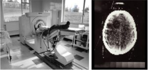
Without strong financial backing, Hounsfield assembled the first machine himself using components from scrap storage. With the quiet support of an executive at EMI, he successfully installed the first scanner at Atkinson Morley Hospital in London. On October 1, 1971, the first CT image of a human brain — revealing a tumor in the left frontal lobe — was produced, marking a historic turning point in modern medicine.
Although EMI did not remain in the medical market for long, Hounsfield’s invention paved the way for major companies such as GE and Siemens to develop full-body CT scanners with greater speed and accuracy. In 1981, he was knighted and continued his research until the end of his life.
From the idea of “seeing through the skull,” CT has now become an indispensable tool in medical diagnosis, especially in emergency medicine, oncology, and trauma care. In 2020, there were more than 80 million CT scans performed in the United States alone. And it all began with a dream — to see the invisible.


Other Posts
.jpg)