

.jpg)

Epidermolysis Bullosa (EB)
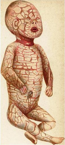
Epidermolysis Bullosa (EB) is a rare group of genetic dermatological conditions characterized by extreme fragility of the skin and mucous membranes. Minor mechanical friction—such as rubbing, gentle pressure, or scratching—causes the skin to tear and form painful blisters. Children born with EB are affectionately referred to as "Butterfly Children" because their skin is as delicate and fragile as a butterfly's wings.
1. Epidemiology and Pathogenesis
-
Incidence: Estimated to affect approximately 1 in 50,000 live births across all subtypes.
-
Genetic Etiology: Caused by mutations in over 18 different genes (such as KRT5, KRT14, LAMA3, LAMB3, LAMC2, and COL7A1). These genes encode structural anchoring proteins (including Keratin, Laminin, and Type VII Collagen) that bind the epidermis (outer layer) to the underlying dermis.
-
Pathophysiology: Deficiency or dysfunction of these structural proteins destroys adhesion between skin layers. Minimal friction causes the epidermis and dermis to separate, allowing fluid accumulation and blister formation.
2. Major Clinical Subtypes
EB is classified into four primary subtypes based on the specific anatomical layer where cleavage and blistering occur:
-
Epidermolysis Bullosa Simplex (EBS):
-
Level of Cleavage: Intraepidermal (within the basal keratinocyte layer).
-
Features: The most common and mildest variant. Blisters primarily affect hands and feet and usually heal without significant scarring.
-
-
Junctional Epidermolysis Bullosa (JEB):
-
Level of Cleavage: Within the lamina lucida of the basement membrane zone.
-
Features: A severe form characterized by widespread blistering, extensive mucosal involvement (oral cavity, larynx, GI tract), and a high rate of infant mortality.
-
-
Dystrophic Epidermolysis Bullosa (DEB):
-
Level of Cleavage: Sub-basal lamina (upper dermis), caused by mutations in the COL7A1 gene encoding Type VII Collagen.
-
Features: Heals with severe scarring, leading to joint contractures, esophageal strictures, and fusion of digits ("mitten" hand deformities).
-
-
Kindler Epidermolysis Bullosa (Kindler Syndrome):
-
Level of Cleavage: Variable (can occur within multiple skin layers).
-
Features: Extremely rare; characterized by photosensitivity, progressive skin atrophy, and poikiloderma.
-
3. Detailed Clinical Presentation
-
Cutaneous Manifestations:
-
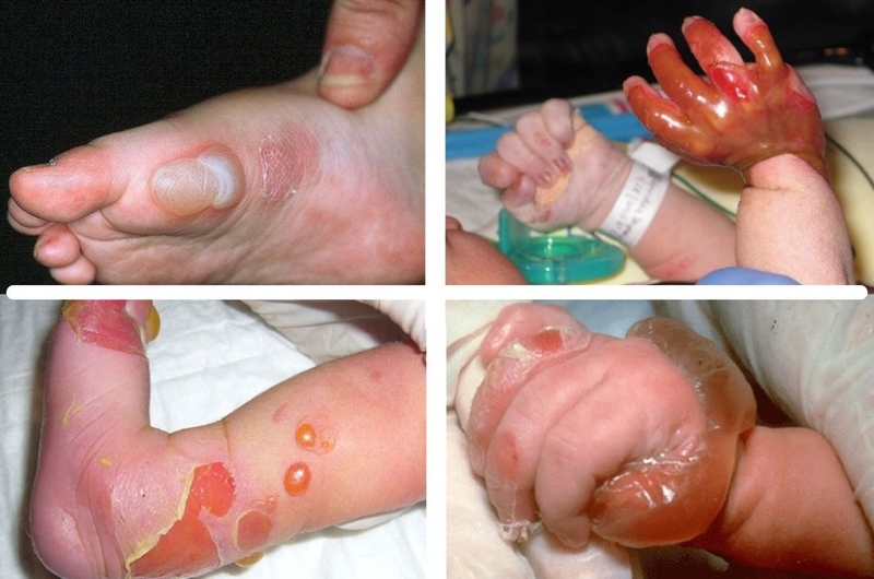
Appearance of clear or hemorrhagic fluid-filled blisters at birth or when an infant begins crawling and walking.
-
Skin tearing, erosions, and extensive sloughing upon mild handling (Nikolsky sign).
-
Nail dystrophy, nail loss, or severe hyperkeratotic nail changes.
-
-
Mucosal and Extracutaneous Involvement:
-
Oral Cavity and Dentition: Painful mucosal ulcerations causing dysphagia, severe early-onset dental caries, and ankyloglossia (tongue-tie).
-
Gastrointestinal Tract: Esophageal blistering and scarring leading to strictures, severe dysphagia, and chronic constipation.
-
Ocular and Respiratory: Corneal erosion leading to visual impairment; airway scarring causing hoarseness or respiratory distress.
-
4. Severe Complications
-
Infection and Sepsis: Extensive denuded skin areas provide a direct portal of entry for pathogens (such as Staphylococcus aureus and Pseudomonas aeruginosa), posing a high risk of life-threatening systemic sepsis.
-
Malnutrition and Chronic Anemia: Caused by painful oral lesions, esophageal strictures, and continuous loss of iron and protein through open blister fluids.
-
Joint Contractures and Disability: Progressive scarring leads to pseudosyndactyly (fusion of fingers and toes) and joint contractures, severely limiting mobility.
-
Squamous Cell Carcinoma (SCC): Patients with severe recessive dystrophic EB (RDEB) face a remarkably high risk of developing aggressive, early-onset skin cancer within chronic scar tissue.
5. Diagnosis and Management
Diagnosis
-
Skin Biopsy (Immunofluorescence Antigen Mapping / Transmission Electron Microscopy): Determines the exact level of tissue cleavage and identifies the missing structural protein.
-
Genetic Testing: Identifies specific gene mutations to confirm subtype, guide prognosis, and offer genetic counseling.
Management and Supportive Care
There is currently no definitive cure for the underlying genetic defect; management focuses on wound care, pain control, and complication prevention:
-
Wound and Skin Care:
-
Lancing new blisters with a sterile needle (while keeping the blister roof intact) to prevent lateral extension of the erosion.
-
Application of specialized non-adherent silicone dressings and gentle wrapping.
-
Complete avoidance of adhesive tapes or bandages directly on the skin.
-
-
Nutritional and Medical Support:
-
High-calorie, high-protein soft or liquid diet.
-
Iron, vitamin, and mineral supplementation. Gastrostomy tube (PEG) placement if esophageal strictures impair oral intake.
-
-
Emerging Therapies: Gene therapy, stem cell transplantation, and topical gene therapy agents (such as Beremagene geperpavec - Vyjuvek) represent breakthrough advancements in restoring functional anchoring proteins.


Other Posts
.jpg)
.jpg)