

.jpg)
Harlequin Ichthyosis
Harlequin Ichthyosis is the most severe and extremely rare genetic form of autosomal recessive congenital ichthyosis. The name "Harlequin" originates from the diamond-patterned costumes of Renaissance traditional jesters, resembling the thick, diamond-shaped skin plates separated by deep fissures that encase the newborn's body.
1. Epidemiology and Incidence
Harlequin Ichthyosis is an extremely rare dermatological genetic disorder:
-
Congenital Incidence: Estimated at approximately 1 in 300,000 to 1 in 500,000 live births.
-
Demographics: It affects males and females equally, with no predilection for specific ethnicities or geographic regions.
-
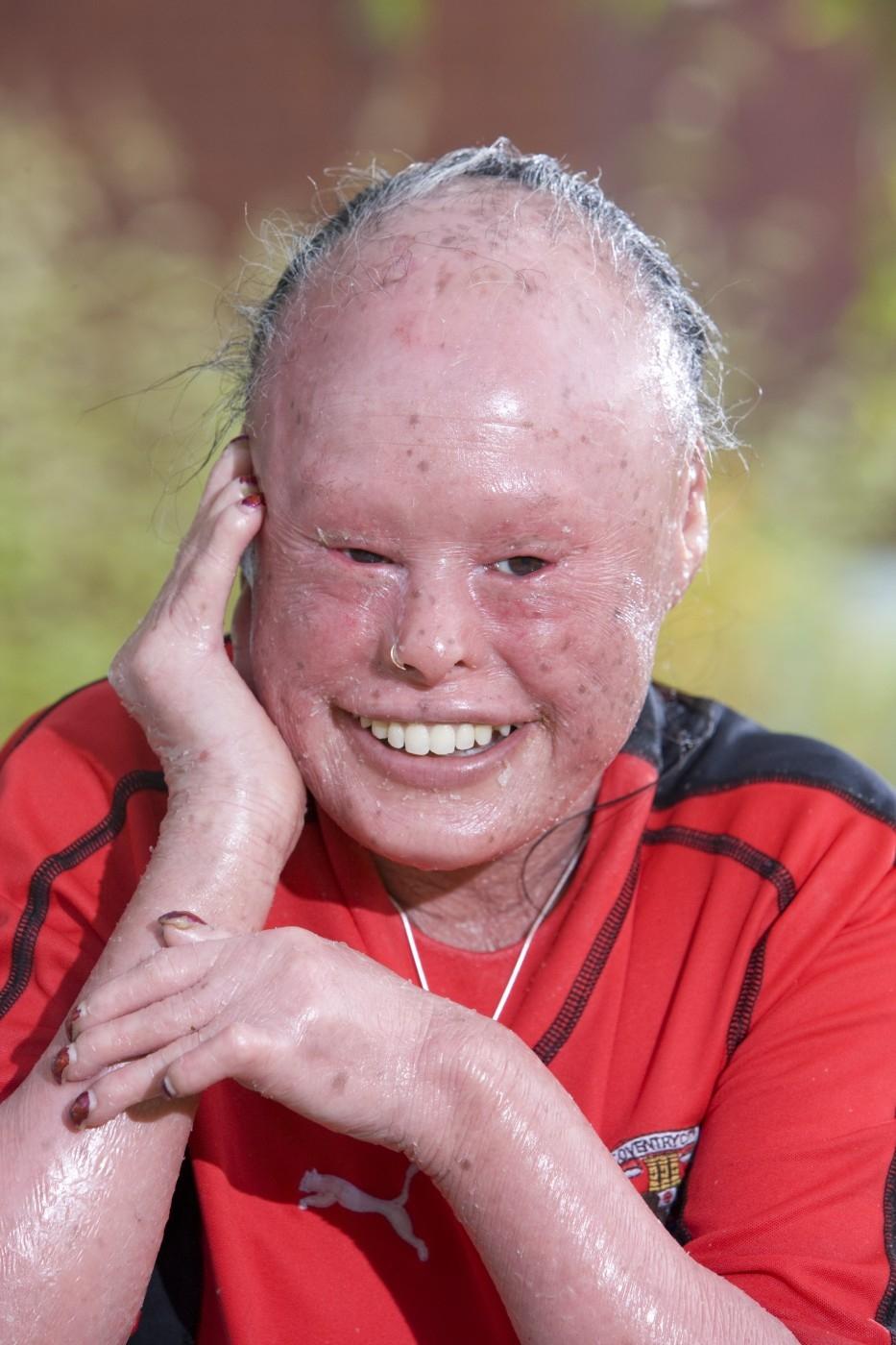
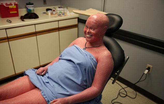
Reported Cases: Historically, high neonatal mortality led to very few survivors; medical literature currently documents a few hundred individuals living with the condition worldwide.
2. Etiology and Genetic Mechanism
ABCA12 Gene Mutation
The condition stems from loss-of-function mutations in the ABCA12 (ATP-binding cassette sub-family A member 12) gene located on chromosome 2 (2q35).
-
Normal Function: The ABCA12 gene encodes a lipid transporter protein responsible for moving lipids into lamellar granules within keratinocytes, essential for skin barrier formation and desquamation.
-
Pathophysiological Consequence: Absence or severe deficiency of the ABCA12 protein impairs lipid transport, disrupting normal skin shedding and causing excessive, thick keratin accumulation (hyperkeratosis).
Autosomal Recessive Inheritance
Harlequin Ichthyosis follows a classic autosomal recessive pattern:
-
An affected infant must inherit two mutated gene copies (one from each parent).
-
Heterozygous parents (carriers) display no clinical symptoms.
-
For each pregnancy of carrier parents, the probabilities are:
-
25% risk of an affected child.
-
50% probability of an asymptomatic carrier child.
-
25% probability of an unaffected, non-carrier child.
-
-
Consanguinity significantly increases the likelihood that both parents carry the rare ABCA12 mutation.
3. Detailed Clinical Presentation
Infants with Harlequin Ichthyosis are frequently born prematurely and exhibit characteristic clinical features at birth.
Armor-Like Hyperkeratotic Plates
The entire body is encased in a thick, rigid layer of hyperkeratotic skin colored grayish-white or yellow. As the infant moves, this rigid armor splits into large polygonal, diamond-shaped plates separated by deep, erythematous fissures that bleed easily.
Facial Deformities
-
Ectropion (Eyelid Eversion): Severe skin tension forces the eyelids to turn inside out, exposing the red inner conjunctival lining. The infant cannot close their eyes, posing a high risk of corneal drying and damage.
-
Eclabium (Lip Eversion): Skin contraction pulls the lips outward, holding the mouth in a fixed, open position.
-
Ear and Nose Deformities: The pinnae (outer ears) and nasal bridge appear flat, underdeveloped, or constricted due to tight skin binding.
Joint and Limb Restrictions
The rigid exoskeleton severely restricts movement, forcing fingers, toes, and limbs into flexed, swollen, or fixed contracture positions.
4. Life-Threatening Complications
The severe loss of normal skin barrier function exposes newborns to critical acute complications:
-
Sepsis: Deep fissures provide a direct portal of entry for pathogens (such as Staphylococcus aureus), leading to severe cutaneous infections and systemic sepsis.
-
Respiratory Distress: The unyielding hyperkeratotic chest wall restricts thoracic expansion, frequently causing respiratory failure.
-
Dehydration and Hypothermia: Excessive transepidermal water loss (TEWL) leads to severe hypernatremic dehydration, electrolyte imbalance, and loss of thermoregulation.
-
Feeding Difficulties: Eclabium prevents effective sucking, necessitating nasogastric or intravenous nutrition.
5. Diagnosis and Management
Diagnosis
-
Prenatal Diagnosis:
-
3D/4D Ultrasound: Identifies structural abnormalities in the 2nd/3rd trimester (fixed open mouth, flexed limbs, skin thickening).
-
Genetic Testing: Amniocentesis or chorionic villus sampling to screen for ABCA12 mutations in at-risk families.
-
-
Postnatal Diagnosis: Confirmed by clinical physical evaluation and molecular genetic verification.
Supportive Treatment Protocol
No curative therapy exists; management focuses on life support and skin barrier maintenance:
-
Neonatal Intensive Care (NICU): Placement in high-humidity incubators (70–80%) to maintain body temperature and reduce skin cracking.
-
Oral Retinoid Therapy: Systemic retinoids (such as Acitretin) accelerate desquamation of thick scales and soften the cutaneous layer.
-
Intensive Emollient Care: Frequent application of thick emollient ointments combined with gentle baths to hydrate skin.
-
Infection Control: Strict sterile environment, topical antiseptics/antibiotics, and IV antibiotics upon sign of infection.
-
Ophthalmological Care: Continuous artificial tears, lubricating eye ointments, and protective moisture chambers to prevent corneal ulceration.
6. Prognosis
-
Historical Context: In the past, most affected infants died within the first few days or weeks of life from sepsis or respiratory failure.
-
Current Status: Advances in NICU supportive care and early systemic retinoid therapy have significantly improved neonatal survival rates. Individuals can reach childhood and adulthood, though they require lifelong intensive dermatological care.


Other Posts
.jpg)
.jpg)




